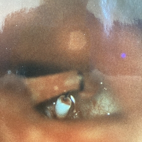
Otosclerosis or Otospongiosis
Otosclerosis is an inheritable genetic disease, transferred by X-chromosome (by women). Otosclerosis aggravation can be triggered by hormonal processes, such as pregnancy.
Otosclerosis is a dysfunction of the ear osseous structure, which occurs in the internal ear (cochlea, vestibule), as well as in the middle ear (the oval window, the stapes and its footplate).
Thus, the ossicular fixation is formed on the level of the junction of the middle and internal ear: on the level of the footplate of stapes.
Beethoven had otosclerosis. During the last years of his life he played the piano posing a stick between his ear and the instrument (without realizing it, he invented a bone conduction hearing device).
The diagnosis is established on the base of clinical examination and audiometry
Clinical examination under microscope will help to ensure the integrity of tympanic membrane.
Otosclerosis is the most common reason of hearing loss (when the tympanic membrane is normal) within young adults.
Acoumetry, a diapason use, will show the hearing loss of transmission and the Bonnier test will be another step towards the confirmation of otosclerosis (you will feel a vibration of diapason posed on the radial or elbow bone).
Audiometry, performed by a hearing specialist, will show the presence of two separated curves on the right and/or on the left (in case of normal hearing these curves form a single line); the absence of acoustic reflex on impedancemetry.
CT-scan of the middle ear is recommended for 3 reasons:
- to exclude pathology with similar clinical signs (the fixed malleus head).
-to exclude abnormalities of anatomical position: facial nerve position in relation with the footplate of stapes, the position of the stapes, abnormality of the auditory canal.
-to exclude thickening of the footplate of stapes.
In case of bilateral otosclerosis, the ear, where the lesion is more pronounced, is operated in the first place.
THE SURGERY
The surgery can be done under local anesthesia (neuroleptanalgesia) or general anesthesia depending on the preferences of the patient. The general anesthesia is used more frequently as it`s more comfortable for the patient.
The surgery is performed through the auditory canal (the endaural approach). The operation can be done fully through the auditory canal with the use of speculum or through a small external incision of Shambaugh in order to broaden the canal, which enables the surgeon to have a better perspective.
The surgery with the use of laser and micro instruments consists of -
-making sure that there is a real ossicular fixation.
-removing the arches of the stapes;
- making a microhole of 6/10 mm on the footplate of the stapes.
-installing a prothesis made of titanium or teflon or a mixed one.
-plugging the zone between the platinotomy and the piston with the conjunctive tissue (a vein graft might be used).
The surgery aims to replace the fixed stapes with a prothesis made of titanium or teflon, 0,4 or 0,6 mm in diameter. A microhole is made through the footplate of the stapes (stapedectomy). The fixed ear bone is removed, the prosthesis is attached to the long limb of the incus.
2 stitches might be made, the eardrum will be held in place by the packing material. A bandage will be placed.
You will be hospitalized for 2 days to ensure the integrity of the inner ear. If after surgery you have vertigo or tinnitus, do not hesitate to inform your doctor – perfusions to protect the inner ear might be necessary. On the contrary, voice resonance is a good sign.
When you are back home, you will be advised to limit your activities – no sport, alcohol, or sauna.
In one week, the bandage will be removed, the packing material will be later absorbed by the body. In a month, you will be recommended to perform an audiometry.
Depending on your lifestyle, you might be advised to use hearing protection.
Swimming will be allowed in a month after surgery, but diving will be prohibited.
You are able to take plane in 2 weeks after surgery.
